Acetabular fossa
| Acetabular fossa | |
|---|---|
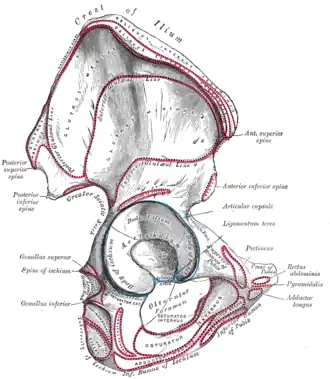 Lateral view of the right hip bone | |
| Details | |
| Identifiers | |
| Latin | fossa acetabuli |
| TA98 | A02.5.01.004 |
| TA2 | 1310 |
| FMA | 17269 |
| Anatomical terms of bone | |
The acetabular fossa is the non-articular depressed region at the centre of the floor of the acetabulum. It is surrounded by the articular lunate surface.[1]: 1368 [2] The floor of the fossa is formed mostly by the ischium;[2] it is rough[1]: 1354 and thin (often to the point of transparency). The space of the fossa is continuous inferiorly with the acetabular notch.[2]
The fossa does not contain any cartilage.[1]: 1368 It is occupied by the ligament of head of femur,[3] and by fibroelastic adipose tissue[4][1]: 1368 (within which the acetabular branch of the obturator artery ramifies[1]: 1250 ) that is mostly lined with synovial membrane.[1]: 1368 The acetabular "fat pad" is thought to contain abundant proprioceptive nerve endings that sense compression of the fat pad or its displacement through the acetabular notch, producing proprioceptive information.[4]
Additional Images
-
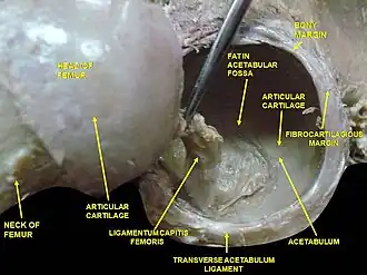 Hip joint. Lateral view. Fat in acetabular fossa.
References
- ^ a b c d e f Standring, Susan (2020). Gray's Anatomy: The Anatomical Basis of Clinical Practice (42th ed.). New York. ISBN 978-0-7020-7707-4. OCLC 1201341621.
{{cite book}}: CS1 maint: location missing publisher (link) - ^ a b c Moore, Keith L.; Dalley, Arthur F.; Agur, Anne M. R. (2018). Clinically Oriented Anatomy (8th ed.). Wolters Kluwer. p. 786. ISBN 978-1-4963-4721-3.
- ^ Tank, Patrick W. (2001). "Bones and Joints of the Pelvis and Perineum - Self Study". University of Arkansas. Archived from the original on 2018-10-12. Retrieved 2008-08-26.
- ^ a b Palastanga, Nigel; Soames, Roger (2012). Anatomy and Human Movement: Structure and Function. Physiotherapy Essentials (6th ed.). Edinburgh: Churchill Livingstone/Elsevier. p. 291. ISBN 978-0-7020-3553-1.