Caldwell's view
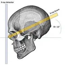
Caldwell's view (or Occipitofrontal view) is a radiographic view of the skull where the X-ray plate is perpendicular to the orbitomeatal line. The rays pass from behind the head and are angled at 15-20° to the radiographic plate. It is commonly used to get better view of the ethmoid and frontal sinuses.[1] It is named after the noted American radiologist Eugene W. Caldwell, who described it in 1907.[2][3]
Structures seen
- Frontal sinus
- Ethmoidal sinus
- Orbit
- Orbital rim
- Medial orbital wall
- Zygomatic bone
- Nasal bone
- Nasal septum
- Mandible
Possible observations
| Pathology | Observation |
|---|---|
| Normal |
|
| Chronic frontal sinusitis |
|
| Osteoma |
|
| Erythroblastic anemia |
|
References
- ^ Yanagisawa, E.; Smith, H. M. (1968-03-01). "Radiographic Anatomy of the Paranasal Sinuses: IV. Caldwell View". Archives of Otolaryngology–Head & Neck Surgery. 87 (3): 311–322. doi:10.1001/archotol.1968.00760060313016. ISSN 0886-4470. PMID 5642379.
- ^ Berk, R N (June 1995). "Eugene W. Caldwell Lecture. Why Caldwell?". American Journal of Roentgenology. 164 (6): 1321–1322. doi:10.2214/ajr.164.6.7754868. ISSN 0361-803X. PMID 7754868.
- ^ Hoeffner, E.G.; Mukherji, S.K.; Srinivasan, A.; Quint, D.J. (December 2012). "Neuroradiology Back to the Future: Head and Neck Imaging". AJNR. American Journal of Neuroradiology. 33 (11): 2026–2032. doi:10.3174/ajnr.A3365. ISSN 0195-6108. PMC 7965588. PMID 23064595.